- Research
- Open access
- Published:
Prognostic value of metabolic dysfunction-associated steatotic liver disease over coronary computed tomography angiography findings: comparison with no-alcoholic fatty liver disease
Cardiovascular Diabetology volume 23, Article number: 167 (2024)
Abstract
Background
Metabolic dysfunction-associated steatotic liver disease (MASLD) is the proposed name change for non-alcoholic fatty liver disease (NAFLD). This study aimed to investigate the association of cardiovascular disease risk with MASLD and NAFLD in patients who underwent clinically indicated coronary computed tomography angiography (CCTA).
Methods
This retrospective study included 2289 patients (60% men; mean age: 68 years) with no history of coronary artery disease who underwent CCTA. The steatotic liver was defined as a hepatic-to-spleen attenuation ratio of < 1.0 on CT just before CCTA. MASLD is defined as the presence of hepatic steatosis along with at least one of the five cardiometabolic risk factors. Adverse CCTA findings were defined as obstructive and/or high-risk plaques. Major adverse cardiac events (MACE) encompassed composite coronary events, including cardiovascular death, acute coronary syndrome, and late coronary revascularization.
Results
MASLD and NAFLD were identified in 415 (18%) and 368 (16%) patients, respectively. Adverse CCTA findings were observed in 40% and 38% of the patients with MASLD and with NAFLD, respectively. Adverse CCTA findings were significantly associated with MASLD (p = 0.007) but not NAFLD (p = 0.253). During a median follow-up of 4.4 years, 102 (4.4%) MACE were observed. MASLD was significantly associated with MACE (hazard ratio 1.82, 95% CI 1.18–2.83, p = 0.007), while its association with NAFLD was not significant (p = 0.070). By incorporating MASLD into a prediction model of MACE, including the risk score and adverse CCTA findings, global chi-squared values significantly increased from 87.0 to 94.1 (p = 0.008).
Conclusions
Patients with MASLD are likely to have a higher risk of cardiovascular disease than those with NAFLD. Concurrent assessment of MASLD during CCTA improves the identification of patients at a higher risk of cardiovascular disease among those with clinically indicated CCTA.
Background
Non-alcoholic fatty liver disease (NAFLD) is a growing public health concern, with an increasing global prevalence of 30% [1]. It is closely associated with obesity and type 2 diabetes [2]. NAFLD is generally considered a hepatic manifestation of metabolic syndrome [3]. Previous studies have demonstrated that NALFD is a significant predictor of cardiovascular disease (CVD) events [4, 5]. International experts have recently published a consensus statement on new fatty liver disease nomenclature, “steatotic liver disease” (SLD) [6]. SLD is classified as metabolic dysfunction-associated SLD (MASLD), MASLD with increased alcohol intake, alcohol-related liver disease, SLD with a specific etiology, and cryptogenic SLD. MASLD is defined as the presence of hepatic steatosis along with at least one of the five cardiometabolic risk factors that correspond to the components of metabolic syndrome [6].
Coronary computed tomography angiography (CCTA) has been established as an accurate diagnostic tool for assessing obstructive and nonobstructive plaque characteristics [7]. Numerous studies have demonstrated the prognostic value of the presence of adverse CCTA findings, defined as obstructive or high-risk plaques, in patients with suspected coronary artery disease (CAD) [8,9,10]. The usefulness of computed tomography (CT) as a measure of SLD has also been reported [11]. Our previous research has demonstrated that NAFLD on nonenhanced CT is significantly associated with the presence of high-risk plaques on CCTA and future CVD events in patients with suspected CAD [4].
The updated diagnostic criteria for MASLD require validation regarding the prediction of CVD risks. This study aimed to clarify additional risk stratification benefits of MASLD or NAFLD concurrently assessed during CCTA in patients with suspected stable CAD in a large cohort.
Methods
Study population
This was a retrospective, single-center cohort study performed at Okayama University Hospital, Japan. Figure 1 shows a flow diagram of the study design. This study enrolled 3570 Japanese outpatients who underwent CCTA between August 2011 and December 2020. Patients with a history of CAD and < 1 year follow-up were excluded. Finally, 2289 patients were included in this study. The study protocol was approved by the Institutional Review Board of Okayama University Hospital, and the study was compliant with the Declaration of Helsinki. Notably, the requirement for informed consent was waived due to the retrospective nature of this study.
Assessment of risk factors
Detailed definitions of risk factors have been described previously [12]. Patients underwent assessments of height, weight, smoking and alcohol history, and other medical histories through physical examination and medical records. Laboratory values, including triglyceride, low-density lipoprotein cholesterol (LDL-C), high-density lipoprotein cholesterol (HDL-C), and hemoglobin A1c levels, were analyzed at the central laboratory of our hospital. Small dense LDL-C levels were calculated using equations reported by Maureen et al. [13]. We calculated that small dense LDL-C = LDL-C– (1.43 × LDL-C– (0.14 × (ln (triglyceride)×LDL-C))-8.99) [13]. The Hisayama risk score (HRS) was used to classify the study population into low- (< 2%), intermediate- (2–10%), and high-risk (> 10%) groups based on the 10-year atherosclerotic CVD risk [14].
Computed tomography assessment of hepatic steatosis
CT scans were performed using a 128-slice CT scanner (SOMATOM Definition Flash; Siemens Medical Solutions, Erlangen, Germany) as previously described [15]. An abdominal non-contrast CT scan was conducted immediately before the cardiac scan on the same day, as previously described [16]. The scan range was 20 cm, and the other scan parameters were 120 kVp, 250 mAs, and 5-mm slice thickness. We used a method for assessing steatotic livers consistent with that of previous reports of the Multi-Ethnic Study of Atherosclerosis [17]. Hepatic and splenic Hounsfield attenuations were measured using the mean Hounsfield unit (HU) in the maximum circular regions of interest (at least 1 cm2) from the two right liver lobes (anteroposterior dimension) and the spleen. The hepatic-to-splenic attenuation ratio was calculated, and a hepatic-to-spleen attenuation ratio of < 1.0 was defined as a positive diagnosis of steatotic liver [11, 17].
Diagnoses of NAFLD and MASLD
MASLD was defined based on the evidence of steatotic liver with the presence of 1 or more of the following five metabolic conditions: (i) body mass index ≥ 23 kg/m2, waist circumference > 94 cm for males and > 80 cm for females or ethnicity adjusted; (ii) fasting serum glucose ≥ 100 mg/dL, 2-hour post-load glucose levels ≥ 140 mg/dL, or hemoglobin A1c ≥ 5.7%, type 2 diabetes, or treatment for type 2 diabetes; (iii) blood pressure ≥ 130/85 mmHg or specific antihypertensive drug treatment; (iv) plasma triglyceride ≥ 150 mg/dL or lipid-lowering treatment; and (v) plasma HDL-C ≤ 40 mg/dL for males and ≤ 50 mg/dL for females or lipid-lowering treatment [6].
NAFLD was defined as the presence of hepatic steatosis without heavy alcohol consumption (ethanol intake > 30 g/day in men and > 20 g/day in women), other coexisting liver diseases such as hepatitis B or C infections, or the use of medications associated with secondary NAFLD (corticosteroids and amiodarone) [18].
Acquisition of CCTA and analyses
Coronary CTA images were obtained as described previously [15]. The acquired data were transferred to a workstation (AZE Virtual Place; Canon Medical Systems Corporation, Otawara, Japan) and reconstructed with a slice thickness of 0.625 mm. During CCTA analysis, we evaluated the degree of stenosis and plaque characteristics in segments with a diameter > 2 mm in accordance with the Society of Cardiovascular Computed Tomography [19]. Plaques were categorized as “calcified” (HU > 130), “non-calcified” (HU < 130), or “low-density” (HU < 50) [15]. Moreover, we defined high-risk plaque (HRP) features (positive remodeling; a remodeling index > 1.1, spotty calcification; a calcium burden length < 1.5, and width less than two-thirds of the vessel diameter, low-density plaque; HU < 30) as previously described [20]. The presence of ≥ 2 features was defined as HRP. Significant stenosis was defined as a luminal narrowing ≥ 50%. Adverse CCTA findings were defined as the presence of significant stenosis and/or HRP. Two experienced cardiovascular imagers (T.N. and T.M.) who were blinded to the clinical data analyzed the CCTA images.
Outcome data
Clinical follow-up was performed by reviewing medical records or telephone interviews. Major adverse cardiac events (MACE) were defined as the composite of cardiovascular death, nonfatal myocardial infarction, and late coronary revascularization. Each outcome was reviewed by clinical event review members (M.N. and T.M.) who were blinded to the CT results according to the relevant criteria. Details of the event definitions are provided in the Additional file. Cardiac death was defined as death due to any of the following causes: acute coronary syndrome (ACS), heart failure, arrhythmic death, or unclear causes of death in which a cardiac origin could not be excluded. ACS includes myocardial infarction and unstable angina. Late coronary revascularization was defined as planned percutaneous coronary intervention or coronary artery bypass grafting due to stable CAD with a new positive functional test for ischemia > 90 days after coronary CTA. MACE occurrence in patients with revascularization scheduled within 90 days on indexed coronary CT findings was excluded to eliminate confounding factors, and these patients were censored at the time of the first revascularization.
Statistical analysis
Continuous variables are expressed as mean ± standard deviation or median with interquartile range. Categorical variables are presented as counts (n) and percentages (%). Continuous variables were compared using the paired Student’s t-test or Mann–Whitney U-test, whereas categorical variables were compared using chi-squared (χ2) analysis or Fisher’s exact test. Cumulative survival estimates were calculated using the Kaplan–Meier method and compared using the log-rank test. The Kaplan–Meier method was applied after categorizing the participants into four groups based on the presence of MASLD or NAFLD and adverse CCTA findings. We performed univariate and multivariate logistic regression analysis to evaluate determinants of adverse CCTA findings, and the results are presented as odds ratios (ORs) with 95% confidence intervals (CIs). The multivariate logistic regression model included age, sex, chronic kidney disease (CKD), current smoking status, and low-density LDL-C. Statin use was also included as a variable. To avoid overlap with the MASLD definition, body mass index, hypertension, dyslipidemia and type 2 diabetes were excluded. To investigate the association of MASLD and NAFLD with MACE, we conducted univariate and multivariate Cox regression analyses, and the results are presented as hazard ratios (HRs) with 95% CIs. The multivariate Cox regression model included the same variables as the multivariate logistic regression model and adverse CT findings. The Hisayama risk score was excluded to avoid overlap with factors in the multivariate model. In the Cox regression model, time was defined as the duration from the baseline to the occurrence of an event or the end of the follow-up period. Furthermore, we assessed the additional predictive value of the presence of MASLD and NAFLD in comparison to adverse CCTA findings for predicting MACE using the global χ2 test. A p-value < 0.05 was considered statistically significant. All statistical analyses were performed using SPSS software (version 29; IBM Corp., Armonk, NY, USA) and the R statistical package (version 4.1.1; R Foundation for Statistical Computing, Vienna, Austria).
Results
Patient characteristics
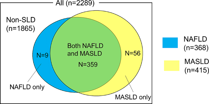
The mean age of the study population was 68 years, and 1371 (60%) patients were men. Among 2289 patients included in the study, 415 (18%) and 368 (16%) were diagnosed with MASLD and NAFLD, respectively. Using the new definition, 56 (2.4%) patients previously not classified as having NAFLD were newly identified as having MASLD (MASLD only) (Fig. 2). Conversely, 9 (0.4%) patients who had been previously classified as having NAFLD did not meet the MASLD criteria (NAFLD only). The remaining 359 (15.6%) patients met both MASLD and NAFLD criteria.
Baseline characteristics of the patients were comparable between those with MASLD and those with NAFLD (Table 1). Patients with MASLD or NAFLD were more likely to be young, male, and to have a higher body mass index, hypertension, dyslipidemia, type 2 diabetes, and CKD than those without MASLD or NAFLD. Additionally, lipid profiles (triglyceride, total cholesterol, HDL-C, LDL-C, small dense LDL-C), AST, and ALT in patients with MASLD or NAFLD were worse than those in patients without MASLD or NAFLD. However, Patients with MASLD were more likely to have elevated HRS compared with those with NAFLD.
Plaque characteristics of MASLD and NAFLD
Plaque characteristics were compared between patients with and without MASLD and between patients with and without NAFLD. As shown in Table 1, patients with both MASLD and NAFLD had a significantly higher prevalence of HRP than those without MASLD and NAFLD (p = 0.001 and p = 0.008, respectively). However, a significant difference in the prevalence of adverse CT findings was observed between patients with and without MASLD rather than between patients with and without NAFLD (p = 0.042 and p = 0.253, respectively).
In Table 2, logistic regression analysis was performed to evaluate determinants of adverse CCTA findings. In the univariate logistic regression analysis, adverse CCTA findings were associated with MASLD (p = 0.039) rather than NAFLD (p = 0.253). Moreover, in the multivariable logistic regression analysis, including variables (age, sex, CKD, current smoking status, statin use, and small dense LDL-C), the association between adverse CCTA findings and MASLD remained significant (p = 0.042).
Association of MASLD and NAFLD with MACE
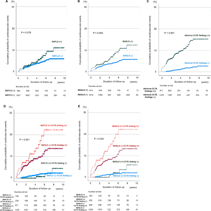
Overall, 102 CVD events were documented during a median follow-up of 4.4 years. Among these, 28 events occurred in patients with MASLD, comprising 3 cardiovascular deaths, 8 myocardial infarctions, and 17 late revascularizations; and 74 events in patients without MASLD: 13 cardiovascular deaths, 13 myocardial infarctions, and 48 late revascularizations). Furthermore, 23 events were observed in patients with NAFLD as follows: 3 cardiovascular deaths, 7 myocardial infarctions, and 13 late revascularization; and 79 events in patients without NAFLD as follows: 13 cardiovascular deaths, 14 myocardial infarctions, and 52 late revascularization. When all participants were categorized according to the presence of MASLD or NAFLD, Kaplan–Meier curves showed that patients with MASLD had higher event rates than patients without MASLD but not NAFLD (Fig. 3A and B; log-rank test, p = 0.003 and p = 0.076). When all participants were categorized according to the presence of adverse CCTA findings, the Kaplan–Meier curves showed that patients with adverse CCTA findings had higher event rates than those without adverse CCTA findings in Fig. 3C (log-rank test, p < 0.001). When all participants were categorized according to the combination of MASLD or NAFLD and adverse CCTA findings, Kaplan–Meier curves showed that patients with both MASLD or NAFLD and adverse CCTA findings had the highest event rates compared to patients without MASLD or NAFLD and adverse CCTA findings (Fig. 3D and E; log-rank test, p < 0.001).
Kaplan–Meier curves stratified according to NAFLD, MASLD, and adverse CCTA findings for MACE. The incidence of MACE during follow-up according to the presence or absence of NAFLD (A), the presence or absence of MASLD (B), the presence or absence of adverse CCTA findings (C), a combination of NAFLD and adverse CCTA findings (D), and a combination of MASLD and adverse CCTA findings (E) CCTA, coronary computed tomography angiography; MASLD, metabolic dysfunction-associated steatotic liver disease; NAFLD, non-alcoholic fatty liver disease
As shown in Table 3, univariate Cox regression analysis showed that MASLD was associated with MACE. Furthermore, in the multivariate Cox regression analysis adjusted for age, sex, CKD, current smoking status, statin use, small dense LDL-C, and adverse CCTA findings, the presence of MASLD was associated with MACE (p = 0.008). However, the presence of NAFLD was not significantly associated with MACE (p = 0.065).
Comparison of predictive performances for MACE
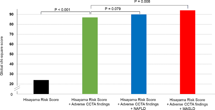
Finally, we assessed whether the inclusion of MASLD or NAFLD to adverse CCTA findings and HRS improved the risk stratification for MACE. Figure 4 illustrates the incremental value of adverse CCTA findings and MASLD or NAFLD in predicting MACE. By considering MASLD along with adverse CCTA findings and HRS, the global χ2 value significantly increased from 87.0 to 94.1 (p = 0.008), while not in NAFLD (p = 0.079). The net reclassification index achieved by incorporating MASLD to adverse CTA findings and HRS was 0.236 (95% confidence interval 0.056–0.415, p = 0.010), while that achieved by adding NAFLD was 0.135 (-0.02 to 0.300, p = 0.107).
The incremental predictive value of NAFLD or MASLD and adverse CT findings and the HRS. A global χ 2 test was used to evaluate the model fitness through adding NAFLD or MASLD for the prediction of MACE in relation to a model of adverse CCTA finding and the Hisayama risk score. CCTA, coronary computed tomography angiography; HRS, Hisayama risk score; MASLD, metabolic dysfunction-associated steatotic liver disease; NAFLD, non-alcoholic fatty liver disease
Discussion
This study demonstrated that MASLD, which was associated with adverse CCTA findings defined as obstructive stenosis and/or HRP, was associated with a higher risk of MACE than NAFLD. Moreover, the presence of MASLD, concurrently assessed during CCTA, along with adverse CCTA findings, enhanced the risk prediction of MACE in patients with clinically indicated CCTA.
To date, no study has reported an increased risk of CVD events in patients with metabolic dysfunction-associated fatty liver disease (MAFLD) compared to those with NAFLD. Previous studies have shown that the higher the number of metabolic components present in individuals with NAFLD, the higher the risk of mortality, highlighting the important roles of metabolic factors in the natural history of NAFLD [21, 22]. In 2020, a new concept called MAFLD was proposed [23]. MAFLD is diagnosed when liver steatosis is present in individuals who are overweight or obese, have type 2 diabetes, or exhibit at least two metabolic risk abnormalities [23]. Although variance between MASLD and MAFLD is anticipated, several studies have reported that MAFLD predicts the risk of CVD events better than NAFLD [24, 25]. The findings of our study are consistent with the importance of metabolic components in cardiovascular outcomes in patients with SLD. The criteria for MASLD include one or more of five cardiometabolic risk factors, thus enabling the identification of patients at a higher risk of CVD.
NAFLD and CVD both share several common metabolic risk factors such as genetics, systemic inflammation, endothelial dysfunction, hepatic insulin resistance, adipose tissue dysfunction, oxidative stress, and lipid metabolism [26, 27]. Moreover, NAFLD is closely linked with various metabolic conditions, which predispose individuals to an elevated risk of CVD [28, 29]. As a result, the patients with NAFLD have tendency to change the composition of serum lipoproteins like smaller peak diameter and particle size and higher particle concentration of LDL-C [30], which was consistent with the result in the present study.
This study revealed that MASLD was more useful than NAFLD in predicting CVD events. There are several possible explanations for these results. First, adverse CCTA findings, including high-risk plaques and significant stenosis, were significantly associated with rather than NAFLD. As shown in this study, adverse CCTA findings significantly affected the incidence of CVD events. Moreover, in this study, patients with MASLD were likely to have a greater high-risk group for HRS than those with NAFLD (22% vs. 19%, respectively). HRS is a risk prediction model for the development of atherosclerotic CVD in Japanese adults [14]. The inclusion criteria for MASLD may have facilitated the identification of the high-risk group for CVD more accurately than those for NAFLD.
This study demonstrated that MASLD concurrently assessed during CCTA significantly improved CVD risk stratification. Performing early and accurate MASLD assessments during CVD risk assessment is crucial. In clinical practice, ultrasonography is typically used to diagnose fatty infiltration; however, non-contrast CT is a useful method for diagnosing liver fat with wide generalization [11]. Based on our findings, utilizing this approach in comprehensive CCTA can enhance the risk stratification of CVD.
Currently, there are no approved medical treatments for MASLD. The primary treatment comprises weight loss through lifestyle interventions, similar to the approach used for NAFLD [31, 32]. Diet and exercise have been found to improve histology, with a greater reduction in inflammation and fibrosis [33]. In patients with type 2 diabetes, pioglitazone, glucagon-like peptide-1 receptor agonists, and sodium glucose cotransporter 2 inhibitors are recommended to improve liver fibrosis [34]. Statins improve cardiovascular outcomes in patients with NAFLD in association with improved aminotransferase levels [35, 36]. Pemafibrate therapy improves markers of hepatic inflammation and fibrosis, regardless of body mass index [37]. These drugs may be effective in improving the prognosis of patients with MASLD. Further studies are required to restore the steatotic liver and interrupt inflammatory and fibrogenic processes.
This study has some limitations. First, the study population was comprised solely by Japanese patients and conducted at a single center. The median age in this study was older than previous studies. Therefore, the results cannot be generalized to other ethnic groups and younger age groups. Second, this study had selection bias because it targeted only patients who underwent clinically indicated CCTA. The prevalence of MASLD (approximately 18% diagnosed using abdominal CT among the enrolled patients) was lower than that reported in previous studies. This discrepancy may be attributed to the differences in the study population, as the enrolled patients in this study, who had clinically indicated CCTA, were different from those in other studies, and the steatotic liver was mostly diagnosed using ultrasonography and magnetic resonance imaging in previous studies. Third, CT results alone may not be sufficient to diagnose SLD, and other examinations other than CT, such as ultrasonography and blood biomarkers, were not performed in our study. Fourth, we did not collect information on changes in medication and risk factor control during the follow-up period, potentially influencing the risk estimates for MASLD. Fifth, our study outlined the feasibility of the simultaneous examination of SLD during CCTA in assessing the risk of cardiovascular events. CCTA is not recommended for a screening of asymptomatic patients. Finally, this was a retrospective observational study. We cannot define a cause-and-effect relationship between MASLD and CVD.
Conclusion
This study demonstrated that the presence of MASLD is significantly associated with MACE and that patients with MASLD may have a higher risk of MACE than those with NAFLD. Moreover, MASLD improved the predictive ability of MACE in addition to adverse CCTA findings in patients who underwent clinically indicated CCTA. Concurrently evaluating MASLD during comprehensive CCTA is effective in identifying patients at a higher risk of CVD events.
Data availability
The datasets used and/or analysed during the current study are available from the corresponding author on reasonable request.
Abbreviations
- CAD:
-
coronary artery disease
- CCTA:
-
coronary computed tomography angiography
- CKD:
-
chronic kidney disease
- CVD:
-
cardiovascular disease
- HDL-C:
-
high-density lipoprotein cholesterol
- HR:
-
hazard ratio
- HRP:
-
high-risk plaque
- HRS:
-
Hisayama risk score
- HU:
-
Hounsfield unit
- MACE:
-
major adverse cardiac events
- MASLD:
-
metabolic dysfunction-associated SLD
- LDL-C:
-
low-density lipoprotein cholesterol
- MAFLD:
-
metabolic dysfunction-associated fatty liver disease
- NAFLD:
-
non-alcoholic fatty liver disease
- SLD:
-
steatotic liver disease
References
Younossi ZM, Golabi P, Paik JM, Henry A, Van Dongen C, Henry L. The global epidemiology of nonalcoholic fatty liver disease (NAFLD) and nonalcoholic steatohepatitis (NASH): a systematic review. Hepatology. 2023;77:1335–47.
Younossi ZM, Henry L. The impact of obesity and type 2 diabetes on chronic liver disease. Am J Gastroenterol. 2019;114:1714–5.
Medina-Santillán R, López-Velázquez JA, Chávez-Tapia N, Torres-Villalobos G, Uribe M. Méndez-Sánchez N. hepatic manifestations of metabolic syndrome. Diabetes Metab Res Rev. 2013. https://doi.org/10.1002/dmrr.2410.
Ichikawa K, Miyoshi T, Osawa K, Miki T, Toda H, Ejiri K, et al. Incremental prognostic value of non-alcoholic fatty liver disease over coronary computed tomography angiography findings in patients with suspected coronary artery disease. Eur J Prev Cardiol. 2022;28:2059–66.
Alon L, Corica B, Raparelli V, Cangemi R, Basili S, Proietti M, et al. Risk of cardiovascular events in patients with non-alcoholic fatty liver disease: a systematic review and meta-analysis. Eur J Prev Cardiol. 2022;29:938–46.
Rinella ME, Lazarus JV, Ratziu V, Francque SM, Sanyal AJ, Kanwal F, et al. A multisociety Delphi consensus statement on new fatty liver disease nomenclature. Hepatology. 2023;78:1966–86.
Miller JM, Rochitte CE, Dewey M, Arbab-Zadeh A, Niinuma H, Gottlieb I, et al. Diagnostic performance of coronary angiography by 64-row CT. N Engl J Med. 2008;359:2324–36.
Motoyama S, Ito H, Sarai M, Kondo T, Kawai H, Nagahara Y, et al. Plaque characterization by coronary computed tomography angiography and the likelihood of acute coronary events in mid-term follow-up. J Am Coll Cardiol. 2015;66:337–46.
Hoffmann U, Ferencik M, Udelson JE, Picard MH, Truong QA, Patel MR, et al. Prognostic value of noninvasive cardiovascular testing in patients with stable chest pain: insights from the PROMISE trial (prospective Multicenter Imaging Study for evaluation of chest Pain). Circulation. 2017;135:2320–32.
Ferencik M, Mayrhofer T, Bittner DO, Emami H, Puchner SB, Lu MT, et al. Use of high-risk coronary atherosclerotic plaque detection for risk stratification of patients with stable chest pain: a secondary analysis of the PROMISE randomized clinical trial. JAMA Cardiol. 2018;3:144–52.
Zeb I, Li D, Nasir K, Katz R, Larijani VN, Budoff MJ. Computed tomography scans in the evaluation of fatty liver disease in a population based study: the multi-ethnic study of atherosclerosis. Acad Radiol. 2012;19:811–18.
Miki T, Miyoshi T, Kotani K, Kohno K, Asonuma H, Sakuragi S, et al. Decrease in oxidized high-density lipoprotein is associated with slowed progression of coronary artery calcification: Subanalysis of a prospective multicenter study. Atherosclerosis. 2019;283:1–6.
Sampson M, Wolska A, Warnick R, Lucero D, Remaley AT. A new equation based on the standard lipid panel for calculating small dense low-density lipoprotein-cholesterol and its use as a risk-enhancer test. Clin Chem. 2021;67:987–97.
Honda T, Chen S, Hata J, Yoshida D, Hirakawa Y, Furuta Y, et al. Development and validation of a risk prediction model for atherosclerotic cardiovascular disease in Japanese adults: the Hisayama study. J Atheroscler Thromb. 2023;30:1966–7.
Suruga K, Miyoshi T, Kotani K, Ichikawa K, Miki T, Osawa K, et al. Higher oxidized high-density lipoprotein to apolipoprotein A-I ratio is associated with high-risk coronary plaque characteristics determined by CT angiography. Int J Cardiol. 2021;324:193–8.
Ichikawa K, Miyoshi T, Osawa K, Miki T, Nakamura K, Ito H. Prognostic value of coronary computed tomographic angiography in patients with nonalcoholic fatty liver disease. JACC Cardiovasc Imaging. 2020;13:1628–30.
Tota-Maharaj R, Blaha MJ, Zeb I, Katz R, Blankstein R, Blumenthal RS, et al. Ethnic and sex differences in fatty liver on cardiac computed tomography: the multi-ethnic study of atherosclerosis. Mayo Clin Proc. 2014;89:493–503.
EASL-EASD-EASO. Clinical practice guidelines for the management of non-alcoholic fatty liver disease. J Hepatol. 2016;64:1388–402.
Shaw LJ, Blankstein R, Bax JJ, Ferencik M, Bittencourt MS, Min JK, et al. Society of Cardiovascular Computed Tomography / North American Society of Cardiovascular Imaging - Expert Consensus Document on coronary CT imaging of atherosclerotic plaque. J Cardiovasc Comput Tomogr. 2021;15:93–109.
Puchner SB, Liu T, Mayrhofer T, Truong QA, Lee H, Fleg JL, et al. High-risk plaque detected on coronary CT angiography predicts acute coronary syndromes independent of significant stenosis in acute chest pain: results from the ROMICAT-II trial. J Am Coll Cardiol. 2014;64:684–92.
Stepanova M, Rafiq N, Younossi ZM. Components of metabolic syndrome are independent predictors of mortality in patients with chronic liver disease: a population-based study. Gut. 2010;59:1410–15.
Golabi P, Otgonsuren M, de Avila L, Sayiner M, Rafiq N, Younossi ZM. Components of metabolic syndrome increase the risk of mortality in nonalcoholic fatty liver disease (NAFLD). Med (Baltim). 2018;97:e0214.
Eslam M, Newsome PN, Sarin SK, Anstee QM, Targher G, Romero-Gomez M, et al. A new definition for metabolic dysfunction-associated fatty liver disease: an international expert consensus statement. J Hepatol. 2020;73:202–9.
Kim H, Lee CJ, Ahn SH, Lee KS, Lee BK, Baik SJ, et al. MAFLD predicts the risk of cardiovascular disease better than NAFLD in asymptomatic subjects with health check-ups. Dig Dis Sci. 2022;67:4919–28.
Chun HS, Lee M, Lee JS, Lee HW, Kim BK, Park JY, et al. Metabolic dysfunction associated fatty liver disease identifies subjects with cardiovascular risk better than non-alcoholic fatty liver disease. Liver Int. 2023;43:608–25.
Ismaiel A, Dumitraşcu DL. Cardiovascular risk in fatty liver disease: the liver-heart axis-literature review. Front Med (Lausanne). 2019;6:202.
Galiero R, Caturano A, Vetrano E, Cesaro A, Rinaldi L, Salvatore T, et al. Pathophysiological mechanisms and clinical evidence of relationship between nonalcoholic fatty liver disease (NAFLD) and cardiovascular disease. Rev Cardiovasc Med. 2021;22:755–68.
Adams LA, Anstee QM, Tilg H, Targher G. Non-alcoholic fatty liver disease and its relationship with cardiovascular disease and other extrahepatic diseases. Gut. 2017;66:1138–53.
Anstee QM, Mantovani A, Tilg H, Targher G. Risk of cardiomyopathy and cardiac arrhythmias in patients with nonalcoholic fatty liver disease. Nat Rev Gastroenterol Hepatol. 2018;15:425–39.
Corey KE, Misdraji J, Gelrud L, Zheng H, Chung RT, Krauss RM. Nonalcoholic steatohepatitis is associated with an atherogenic lipoprotein subfraction profile. Lipids Health Dis. 2014;13:100.
Romero-Gómez M, Zelber-Sagi S, Trenell M. Treatment of NAFLD with diet, physical activity and exercise. J Hepatol. 2017;67:829–46.
Ampuero J, Sánchez-Torrijos Y, Aguilera V, Bellido F, Romero-Gómez M. New therapeutic perspectives in non-alcoholic steatohepatitis. Gastroenterol Hepatol. 2018;41:128–42.
Vilar-Gomez E, Martinez-Perez Y, Calzadilla-Bertot L, Torres-Gonzalez A, Gra-Oramas B, Gonzalez-Fabian L, et al. Weight loss through lifestyle modification significantly reduces features of nonalcoholic steatohepatitis. Gastroenterology. 2015;149:367–78..e365; quiz e314-365.
Cusi K, Isaacs S, Barb D, Basu R, Caprio S, Garvey WT, et al. American Association of Clinical Endocrinology Clinical Practice Guideline for the diagnosis and management of nonalcoholic fatty liver Disease in Primary Care and Endocrinology Clinical Settings: co-sponsored by the American Association for the study of Liver diseases (AASLD). Endocr Pract. 2022;28:528–62.
Athyros VG, Tziomalos K, Gossios TD, Griva T, Anagnostis P, Kargiotis K, et al. Safety and efficacy of long-term statin treatment for cardiovascular events in patients with coronary heart disease and abnormal liver tests in the Greek Atorvastatin and Coronary Heart Disease evaluation (GREACE) study: a post-hoc analysis. Lancet. 2010;376:1916–22.
Tikkanen MJ, Fayyad R, Faergeman O, Olsson AG, Wun CC, Laskey R, et al. Effect of intensive lipid lowering with atorvastatin on cardiovascular outcomes in coronary heart disease patients with mild-to-moderate baseline elevations in alanine aminotransferase levels. Int J Cardiol. 2013;168:3846–52.
Shinozaki S, Tahara T, Miura K, Lefor AK, Yamamoto H. Pemafibrate therapy for non-alcoholic fatty liver disease is more effective in lean patients than obese patients. Clin Exp Hepatol. 2022;8:278–83.
Acknowledgements
None.
Funding
This study was supported by the Japan Society for the Promotion of Science KAKENHI (21K08052).
Author information
Authors and Affiliations
Contributions
T.N. contributed to conceptualization, investigation, data collection and validation, supervision, formal analysis, and original draft writing. T.M. contributed to conceptualization, methodology, formal analysis, and reviewing and editing. M.N., T.M., H.T., M.K., K.I., K.O. contributed to investigation, data collection, and reviewing and editing. S.Y.contributed to supervision, and reviewing and editing.
Corresponding author
Ethics declarations
Ethics approval and consent to participate
The study protocol was approved by the Institutional Review Board of Okayama University Hospital, and the study conducted in accordance with the Declaration of Helsinki. The requirement for informed consent was waived owing to the study’s retrospective design.
Competing interests
The authors declare no competing interests.
Additional information
Publisher’s Note
Springer Nature remains neutral with regard to jurisdictional claims in published maps and institutional affiliations.
Supplementary Information
Rights and permissions
Open Access This article is licensed under a Creative Commons Attribution 4.0 International License, which permits use, sharing, adaptation, distribution and reproduction in any medium or format, as long as you give appropriate credit to the original author(s) and the source, provide a link to the Creative Commons licence, and indicate if changes were made. The images or other third party material in this article are included in the article’s Creative Commons licence, unless indicated otherwise in a credit line to the material. If material is not included in the article’s Creative Commons licence and your intended use is not permitted by statutory regulation or exceeds the permitted use, you will need to obtain permission directly from the copyright holder. To view a copy of this licence, visit http://creativecommons.org/licenses/by/4.0/. The Creative Commons Public Domain Dedication waiver (http://creativecommons.org/publicdomain/zero/1.0/) applies to the data made available in this article, unless otherwise stated in a credit line to the data.
About this article
Cite this article
Nishihara, T., Miyoshi, T., Nakashima, M. et al. Prognostic value of metabolic dysfunction-associated steatotic liver disease over coronary computed tomography angiography findings: comparison with no-alcoholic fatty liver disease. Cardiovasc Diabetol 23, 167 (2024). https://doi.org/10.1186/s12933-024-02268-1
Received:
Accepted:
Published:
DOI: https://doi.org/10.1186/s12933-024-02268-1


